
Videosomnoscopy (DISE): what it is and why it decides your treatment
Drug-induced sleep endoscopy in Madrid
Medically reviewed by Dr. Iván Méndez-Benegassi Silva · Otorhinolaryngology (ENT) Specialist with over 25 years of experience in sleep surgery and advanced rhinology · European Board Certificate (ORL-HNS) · HM Hospitales, Madrid · License No. 282855946
What videosomnoscopy (DISE) is
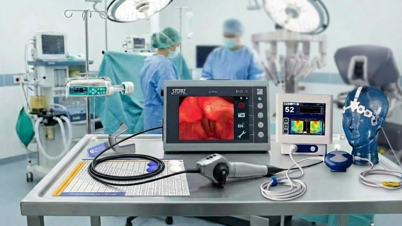
Videosomnoscopy — also called DISE (drug-induced sleep endoscopy) — is a diagnostic test, not an operation. It is performed in people with night-time snoring or sleep apnea. It consists of looking at the upper airway with a flexible endoscope while the patient sleeps a medication-induced sleep, to see with one’s own eyes at which level the throat closes. It is practically the only way to observe the obstruction while the person is asleep (the only alternative, in selected cases, is measuring the pressure inside the pharynx).
Step by step
It is an outpatient procedure performed in an operating room or suite with an anaesthetist. First, the anaesthetist induces a controlled sleep intravenously; it is not general anaesthesia and, under normal conditions, the patient is not intubated (intubation would only be an exceptional rescue measure). Then the otolaryngologist passes a flexible endoscope (a very thin, soft tube) through the nose to the throat and observes the airway while the snoring and breathing pauses are reproduced. Where, how much and how it obstructs is recorded. The examination lasts about 30 minutes and, being outpatient, the patient is discharged the same day.
How sleep is induced: sedation, BIS and TCI pump
Sleep is induced with drugs always administered by the anaesthetist, usually propofol (sometimes dexmedetomidine). To know objectively whether the sedation is at the right point, a BIS monitor (bispectral index) is used: from the brain’s electrical activity — a simplified electroencephalogram (EEG) — it produces a number that indicates the depth of sleep, instead of relying on the subjective impression of the person performing the test. There is an ideal window in which the induced sleep resembles natural sleep; above it the patient is too awake and the apnea is underestimated, below it too deep and it is overestimated. A TCI pump (target-controlled infusion) is also usually used to keep the dose stable so that window does not drift. The aim is to reproduce a sleep as close as possible to sleep at home.
What is seen: the VOTE classification in plain terms
The test identifies the site, the degree and the pattern of collapse, recorded with the VOTE classification (Kezirian, 2011), which looks at four areas: V for velum or soft palate, O for oropharynx (the lateral walls), T for tongue base and E for epiglottis (the flap of cartilage that covers the airway). For each area it notes how much it closes (0 none, 1 partial, 2 fully) and how: anteroposterior (front to back), lateral (side to side) or concentric (in a circle, like a ring). The areas that close most are the palate (80-92%), the tongue base (52-61%), the lateral walls (50-60%) and the epiglottis (30-38%). A key fact: what closes while asleep is different from what is seen awake in around 75% of cases.
Videosomnoscopy step by step (video)
Why it should come BEFORE a mandibular advancement device (MAD)
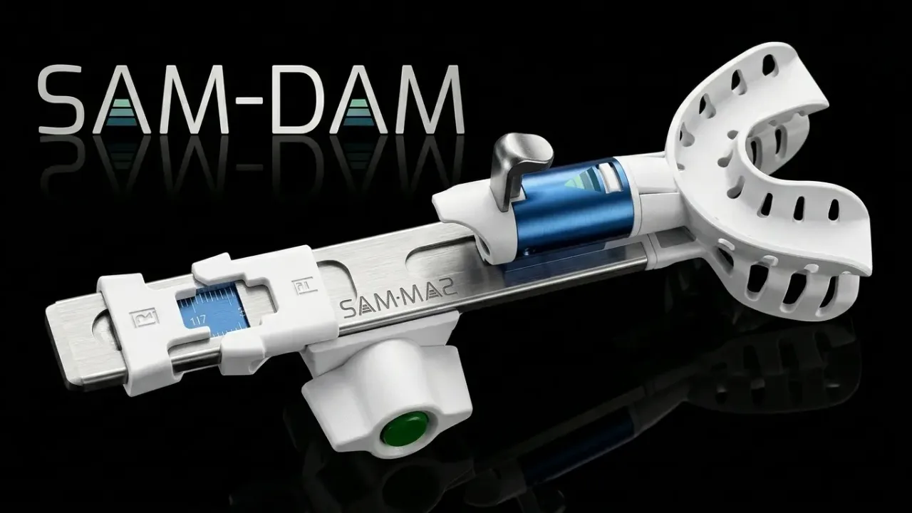
The best way to know whether a mandibular advancement device (MAD) will help a patient is to check it while asleep, because sleep is dynamic: muscle relaxation, inspiratory effort, posture and how the tongue pushes all depend on it. An assessment made while awake does not reflect that. During videosomnoscopy the appliance test is done: a positioner moves the jaw forward in measured, millimetre steps (the DISE-SAM protocol) to check whether the airway opens. What is seen helps to predict the result: collapse at the tongue base predicts a good response to the device (around three times the chance of responding), whereas a complete concentric collapse of the palate (around 5 times the risk of worsening) or a complete lateral collapse of the walls (around 6-7 times) warn that the device may even make things worse. In fact, when the device is fitted after checking it asleep, its success rises to close to 83%, versus 69-75% when fitted without checking. Checking beforehand avoids spending on a quality device that is not going to work; poor-quality devices also fall out, are uncomfortable and do not fit well.
Sleeping on your side may be enough: positional therapy
During the test the patient is observed asleep on their back (supine) and on their side (lateral), because posture changes the obstruction a great deal. Up to half of patients improve simply by sleeping on their side, sometimes without needing any other treatment. This is positional therapy, a simple measure that patients are often not told about. Videosomnoscopy makes it possible to identify who could manage with this very simple measure before considering anything else.
The collapse pattern decides the surgical technique
It is not enough to know where the throat closes: how it closes matters. An anteroposterior collapse (front to back) calls for a different type of pharyngoplasty (surgery on the pharyngeal walls) than a lateral or concentric one, and success rates change with the pattern; choosing the wrong technique worsens the result. The test also detects tongue-palate coupling — whether the tongue pushes against the palate: when this happens, isolated palate surgery would fail, so it is worth knowing before operating.
When the closure is at the palate or the side walls, the operation is a pharyngoplasty (palate surgery), and its type is chosen according to the pattern seen in the DISE.
About pharyngoplastySurgical simulation and multilevel surgery
The most common finding is not a single closure point but multilevel collapse: several areas closing at once, something that can only be seen while the person sleeps (68-76% of patients). During the test, when one collapse is relieved, it is checked whether other obstructions appear lower down that would make single-site surgery fail. This is how it is decided, on solid grounds, whether several areas of the pharynx need operating (multilevel surgery) instead of just one — or even whether it is better not to operate.
If the tongue base also closes, it can be reduced through the mouth with transoral robotic surgery (TORS); the DISE shows whether that level is involved.
Hypoglossal implant (Nyxoah)Locating the origin of the snoring
Beyond apnea, videosomnoscopy makes it possible to determine where the snoring originates in each person — the palate, the pharyngeal walls or the tongue base. Knowing that origin helps to plan, if treatment is chosen, the specific snoring surgery and to avoid operating on areas that are not responsible for the noise.
It unmasks nasal obstruction that only appears during sleep
The videosomnoscopy also reveals nasal obstructions that only occur during sleep. Lying down, many patients develop an obstructive rhinitis that is not apparent during the day and that, being asleep, they are not aware of. That blocked nose at night worsens sleep quality and indirectly forces the mouth open, which increases snoring and makes it harder to adapt to CPAP. Detecting it makes it possible to also treat the nose when it is part of the problem.
If the nose is part of the problem, it is treated with a septoplasty when the septum is deviated or a turbinate reduction (turbinoplasty) when the turbinates are enlarged; if the palate also vibrates, septal graft pharyngoplasty uses the septal cartilage to stiffen it in the same operation. To measure how blocked your nose feels before the consultation, you can fill in the NOSE nasal obstruction questionnaire.
Precise sedation: neither too asleep nor too awake
The balance of sedation is one of the keys to the quality of the test, which is why the BIS monitor and the TCI pump are used to keep it stable. If the patient is too deeply asleep (over-sedation), collapses appear that are not real and one might believe, for example, that a device will not work when in fact it would: a false negative that would deny the patient a valid treatment. If they are too awake, the real collapses stay hidden and it is no use either. The right point is what makes what is observed reliable and reproducible.
Who it is for (and when not)
It is indicated mainly in people with obstructive sleep apnea or snoring who are going to consider surgery or a device, and in those who do not tolerate or refuse CPAP. It is not advisable in every case and has contraindications: pregnancy, allergy to the sedation drugs, high anaesthetic risk or serious clotting problems. In children it is usually not necessary, since the most common cause is the tonsils and adenoids.
If you cannot get used to CPAP, the guide CPAP intolerance: what real alternatives do I have? reviews the options and when each one makes sense.
The patient pathway: who does what
Sleep apnea is first diagnosed by a pulmonologist or a neurophysiologist through the sleep study; CPAP is their reference treatment. When any alternative to CPAP is considered, the patient should be seen by an otolaryngologist, who is the one who determines the treatment or combination of treatments. The dentist —ideally a specialised one, since not all are— does not decide the indication: they make and fit the oral appliance using the measurements obtained during the videosomnoscopy. And if the patient is a good candidate for facial-skeleton surgery (maxillomandibular advancement, maxillary expansion…), the otolaryngologist refers them to the maxillofacial surgeon. In short, the ENT is the reference point for every alternative to CPAP.
Is it safe? Where it is done and its honest limits
Because unstable sedation can be dangerous, the test must be performed in a hospital setting with an anaesthetist and safety standards; it is not an examination to be done just anywhere. Its risks are few: sometimes the snoring cannot be reproduced, secretions may make interpretation difficult, and there may be minor discomfort in the nose or throat. Serious complications are very exceptional; in especially fragile patients, serious situations (breathing difficulty or bleeding) that could require intubation have rarely been described, with a minimal mortality risk — the same warning given in the official consent form. And to be honest: the evidence on whether it improves surgical outcomes is mixed. It is a tool to decide better, not a promise of a result.
SEORL-CCC informed consent for DISE (in Spanish)Where it is done in Madrid
I perform and interpret videosomnoscopy (DISE) personally at HM group hospitals in Madrid, alongside a specialized anaesthesia team with full hospital safeguards. I assess and treat the upper airway in close coordination with the anaesthetist and, when indicated, the sleep pulmonologist.
Consensus document on DISE
I am a co-author of the Spanish national consensus document on drug-induced sleep endoscopy (DISE):
Carrasco Llatas M, Martínez Ruiz de Apodaca P, Baptista Jardín P, O'Connor Reina C, Plaza Mayor G, Méndez-Benegassi Silva I, et al. Drug-induced sleep endoscopy. Acta Otorrinolaringol Esp (Engl Ed). 2020;71(5):316-320. PMID: 31174844 · DOI: 10.1016/j.otorri.2019.02.007
Frequently asked questions
Does the videosomnoscopy hurt?
No. It is a minimally invasive, painless test: the patient is asleep under sedation throughout the examination and does not feel the endoscope. Afterwards, at most, there may be slight nasal or throat discomfort that resolves on its own.
Is the sedation dangerous?
When performed in a hospital setting, with an anaesthetist controlling the depth of sleep with a BIS monitor and an infusion pump, it is a safe test. It is not general anaesthesia and the patient is not intubated. A serious risk is very exceptional and limited to especially fragile patients.
How long does it take and do I need to be admitted?
It is an outpatient procedure: no admission is needed. The examination lasts about 30 minutes. The patient is discharged the same day and should go accompanied and not drive, because of the sedation.
How many days off work?
The videosomnoscopy on its own does not usually need more time off than the day of the test: if no operation is done in the same session, the official consent form of the Spanish ENT society (SEORL-CCC) states that you can go home a few hours later, with the usual advice after an outpatient sedation; that day you should go accompanied and not drive. If the DISE is done just before an operation, in the same operating room, the days off depend on the operation, not on the test. Sick leave is decided by your doctor according to your job.
How is it different from a sleep study (polysomnography)?
The sleep study measures how much apnea there is (the severity), but it does not say where or how the airway collapses. Videosomnoscopy is complementary: it shows at which level of the throat the airway closes while you sleep, and how. One measures the amount of the problem; the other shows its location.
Does it always end in surgery?
No. It serves both to indicate surgery and to rule it out: it can suggest that an oral appliance or CPAP may be enough, show that sleeping on your side could be enough, or avoid an operation that was not going to work. It is a tool to choose the right treatment, whatever that is.
Will I be aware of anything during the test?
No. The patient sleeps under sedation throughout the examination and remembers nothing of it. On waking they are in the recovery area; afterwards the specialist explains the findings and the treatment advised based on what was seen.
When will I have the results?
In the same session. As it is seen live, the specialist knows the site, the degree and the pattern of collapse straight away, and afterwards explains the most suitable treatment for your case.
Take the next step in your evaluation
Dr. Méndez-Benegassi uses your data to answer your message. Email is not end-to-end encrypted: it can be read on the servers it passes through. Do not send medical reports or health data that way.
Data controller: Dr. Iván Méndez-Benegassi Silva, ENT physician.
Purpose: To read and answer your message. Tell us only what is essential; the rest is covered in the consultation.
Legal basis: The healthcare you are requesting (Art. 9.2.h GDPR).
Recipients: Your data is not shared with third parties; only the technical providers needed are involved, as data processors.
Your rights: Access, rectify or erase your data, object, restrict its processing and request its portability by writing to contacto@ronquidoyapneas.com. You may also lodge a complaint with the Spanish Data Protection Agency.
Full details in the Privacy policy.
This guide is educational and does not replace medical advice. The videosomnoscopy (DISE) and its indication must be assessed by a specialist on an individual basis.
Reviewed by Dr. Iván Méndez-Benegassi Silva · Otorhinolaryngology (ENT) Specialist with over 25 years of experience in sleep surgery and advanced rhinology · European Board Certificate (ORL-HNS) · HM Hospitales · Last reviewed: 21 Jun 2026
Where I see patients in Madrid
I see patients in person at my HM Hospitales clinics (HM Madrid, HM Valdebebas and HM Tres Cantos), or by personalized video consultation if you prefer.
Private consultation, no waiting lists
Call the appointments line: you get your appointment during the same call, at any of my three clinics.
Call · 910 786 601- HM Madrid
Wednesdays 3:00 PM - 4:00 PM - HM Valdebebas
Tuesdays 3:00 PM - 4:00 PM - HM Tres Cantos
Tuesdays 1:30 PM - 2:30 PM
€150, paid at the reception desk of the clinic. It is not processed through insurance, except for reimbursement insurance.
Coming with your health insurance? Book with HM: 919 370 000.
HM Madrid
HM Madrid University Hospital
Pl. del Conde del Valle de Súchil, 16
28015 MadridWith insurance (book with HM): Wednesdays 4:00 PM - 7:00 PM
How to get thereHM Valdebebas
HM Valdebebas Polyclinic
Av. de Juan Antonio Samaranch, 51
28055 MadridWith insurance (book with HM): Tuesdays 4:00 PM - 7:00 PM
How to get thereHM Tres Cantos
HM Tres Cantos University Hospital
C. Santiago Ramón y Cajal, 23
28760 Tres CantosWith insurance (book with HM): Tuesdays and Wednesdays 9:30 AM - 1:30 PM
How to get there